Segment VI/VII segmentectomy
Case Scenario
- The patient was a 74 year old woman with Child's A cirrhosis (likely on the basis of NASH) and an asymptomatic 4 cm hepatocellular carcinoma, identified during investigation of an unrelated problem.
- The bilirubin was 5 mmol/L , albumin was 40 g/L, INR was 1.03 and the platelet count was 340.
- The tumour was somewhat exophytic; it lay within segments VI & VII. Contrast-enchanced ultrasound confirmed the solitary lesion.
- Surgical resection is preferred over trans-arterial chemoembolization (TACE), radiation or Sorafenib. In the presence of cirrhosis, a parenchymal sparring procedure is preferred. Based on the relationship with the right hepatic vein, a formal resection of segment VI & VII was planned.
Pathology Slides
- Images courtesy of Dr. Oyedele Adeyi, Toronto General Hospital. Click on the thumbnails below to view the pathology slides in full size. Best viewed in Mozilla Firefox, Google Chrome or Safari.
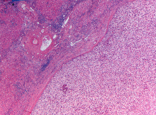
Steatotic tumor (right half) compared to non-steatotic background cirrhosis. (Hematoxylin and Eosin Stain, 25x)
Steatotic tumor (right half) compared to non-steatotic background cirrhosis. (Hematoxylin and Eosin Stain, 25x)
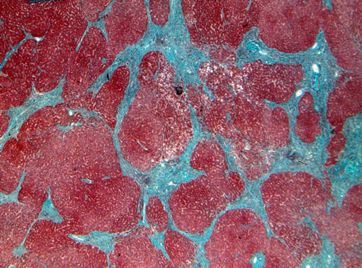
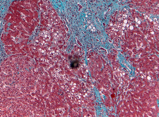
The cirrhotic background is highlighted with little to no evidence of NASH (i.e. burnt-out) - something that not infrequently occurs as cirrhosis advances. (Trichrome stain, 25x)
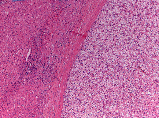
There is little residual steatosis in the background. (Trichrome stain, 100x)
- Images courtesy of Dr. Oyedele Adeyi, Toronto General Hospital. Click on the thumbnails below to view the pathology slides in full size. Best viewed in Mozilla Firefox, Google Chrome or Safari.