Modeling
The patient's actual CT scans are used as a base to build the 3D models. Typically a CT data set with strong contrast would be selected, as structures (vessels, tumors) will appear prominently.
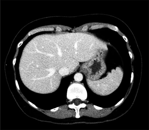
CT scan from patient.
Using the Myrian XP Liver software, a rough model of the structure is created (right). Seen here is a rough model of the hepatic venous system.
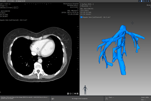
Screenshot of the Myrian XP Liver software.
This rough model is then imported into Cinema 4D, a 3D modeling and animation software package. Notice the ragged edges and artifacts in the rough model.
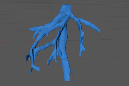
Rough model imported into Cinema4D.
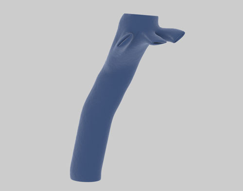
Using the rough model as a reference, a smoothed and simplified model is created, showing only the relevant structures. In this case, numerous small branches were omitted in the simplified model.
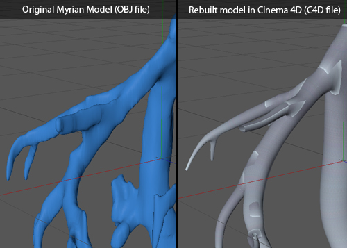
Rebuilding the model.
The final model is then textured and rendered for an aesthetic appearance.
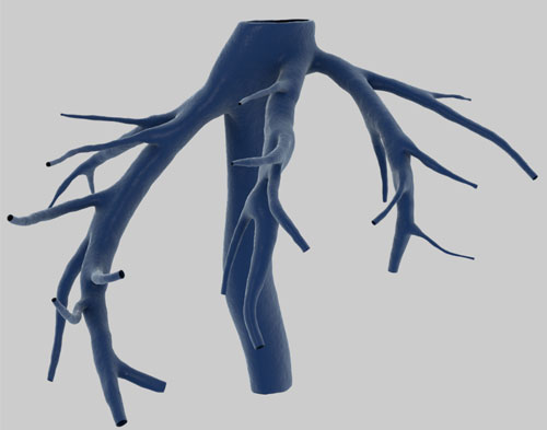
Texturing/rendering the model.
The model can also be modified to fit procedures already performed. In this case, all 3 hepatic branches have been divided in the surgery, and this is shown in the model.
Modifying the model to fit surgical procedures.
Animation
A freeze frame is then taken from the surgical video footage. These shots are typically staged, to show the relevant anatomy with minimum obstruction.
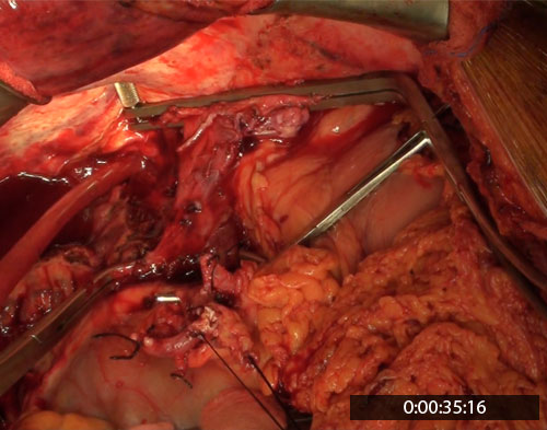
Taking a freeze frame in the surgical video.
The model can then be matched exactly to the structure seen in the freeze frame.
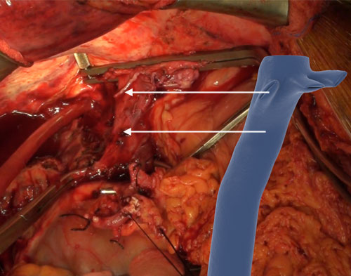
Matching the 3D model to the frozen frame.
This allows a smooth, accurate transition between surgical video and animation, which is the key to our approach in enhancing the educational value in surgical videos.
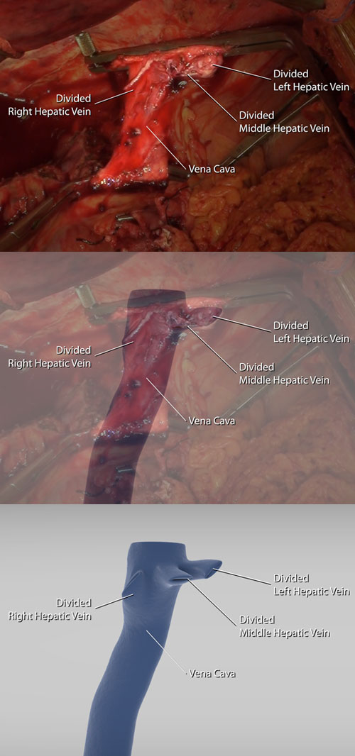
Transition from surgical video to animation.
We then utilize animation to convey important educational messages. In this case, we show the proposed venotomy in the vena cava vessel wall.
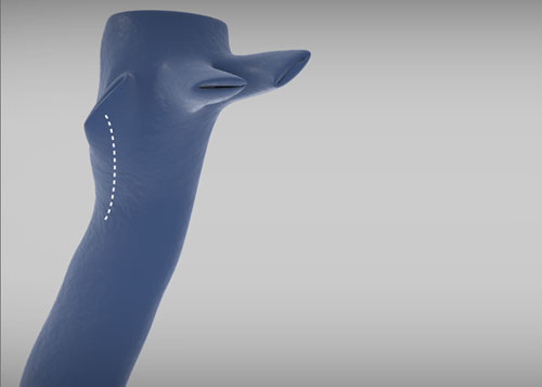
Animation showing further details of the procedures to be performed.