
Content
Focused Cardiac Ultrasound (FOCUS)
FOCUS Cardiac Ultrasound Course
![]() These 1-day courses are designed to introduce physicians involved in the acute care of patients to the theory and practice of Focused Cardiac Ultrasound (FOCUS). The courses will take place on Jan 24, Mar 7 and June 6, 2015. Download the course brochure.
These 1-day courses are designed to introduce physicians involved in the acute care of patients to the theory and practice of Focused Cardiac Ultrasound (FOCUS). The courses will take place on Jan 24, Mar 7 and June 6, 2015. Download the course brochure.
Page Content for the FOCUS online module:
Access the Module
The TTE FOCUS module now contains a section covering cardiac pathology. The first condition covered in this section is pericardial effusion, with examples of a localized pericardial effusion and tamponade. More pathological conditions that can be assessed with FOCUS will be added in the coming months.
Click here to open the Focused Cardiac Ultrasound application.
Using an iPad? Click the App Store button to buy the app.
Updating the TTE FOCUS Views app:
Users who are updating the TTE FOCUS Views app from the previous version, please delete the app from your iPad and then reinstall the new version from your purchased apps in the App Store. If you simply update the app, there is a problem accessing the normal TTE views. We apologize for the inconvenience, and are working on a solution for this problem.
Introduction
In the practice of anesthesia, critical care and emergency medicine there is often a need for a quick, qualitative assessment of cardiac function. Focused Cardiac Ultrasound (FOCUS) is the process of carrying out this rapid qualitative assessment by practitioners in these fields. Training for carrying out a FOCUS assessment requires practitioners to understand the 3D structures of the heart that are seen in the 2D TTE image.
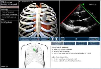
We have created this online interactive module to assist with teaching and learning the assessment of cardiac function with FOCUS. Users can view the TTE recordings for each of the 5 FOCUS views and see a corresponding 3D model of the probe, ultrasound plane, heart and rib cage for each view.
FOCUS assessments based on a subset of 4 of the 20 standard transthoracic echocardiography (TTE) views, with the addition of an inferior vena caval view that is not part of a standard TTE exam.
- Parasternal long axis
- Parasternal short axis
- Apical four chamber
- Subcostal four chamber
- Subcostal inferior vena cava
Instructions
Each FOCUS view can be selected from a drop-down menu in the upper left of the screen. For each FOCUS view, the 3D model of the probe, ultrasound plane, heart and rib cage can be rotated in the horizontal or vertical plane to view it from any angle.
The rib cage can be removed, the part of the heart above the echo plane can be removed, and the heart model can be oriented so the structures correspond to the TTE image.