The Toronto Video Atlas of Liver, Pancreas and Transplant Surgery is happy to announce our publication in the Journal of Visualized Surgery.
In collaboration with Dr.s Anne Agur and Jodie Jenkinson of the University of Toronto, "Depicting surgical anatomy of the porta hepatis in living donor liver transplantation" describes the process of developing an animation-enhanced surgical video based on live OR footage and patient-specific 3D anatomical models.

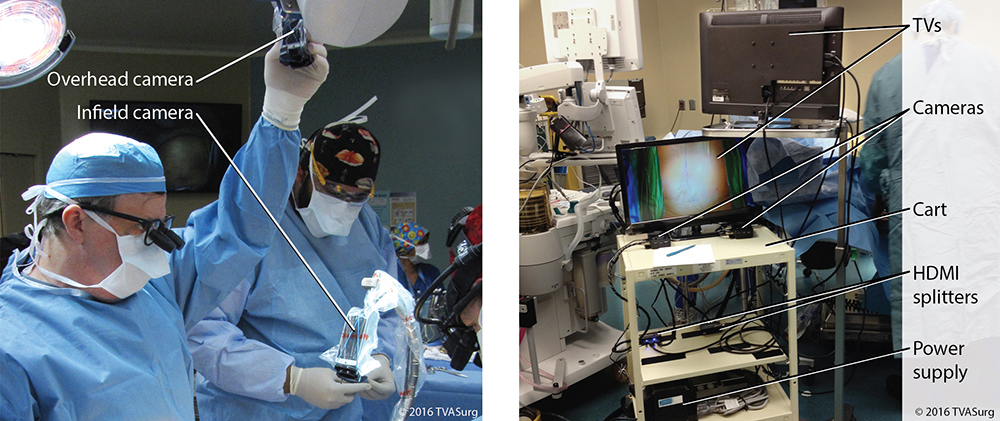
Figure 1 from the publication: A) Overhead and infield cameras in the operating room. B) The filming cart used by the medical illustrators.
ABSTRACT
BACKGROUND: Visualizing the complex anatomy of vascular and biliary structures of the liver on a case-by-case basis has been challenging. A living donor liver transplant (LDLT) right hepatectomy case, with focus on the porta hepatis, was used to demonstrate an innovative method to visualize anatomy with the purpose of refining preoperative planning and teaching of complex surgical procedures.
METHODS: The production of an animation-enhanced video consisted of many stages including the integration of pre-surgical planning, case-specific footage and 3D models of the liver and associated vasculature, reconstructed from contrast-enhanced CTs. Reconstructions of the biliary system were modeled from intraoperative cholangiograms. The distribution of the donor portal veins, hepatic arteries and bile ducts was defined from the porta hepatis intrahepatically to the point of surgical division. Each step of the surgery was enhanced with 3D animation to provide sequential and seamless visualization from pre-surgical planning to outcome. Use of visualization techniques such as transparency and overlays allows viewers not only to see the operative field, but also the origin and course of segmental branches and their spatial relationships.
RESULTS: This novel educational approach enables integrating case-based operative footage with advanced editing techniques for visualizing not only the surgical procedure, but also complex anatomy such as vascular and biliary structures. The surgical team has found this approach to be beneficial for preoperative planning and clinical teaching, especially for complex cases. Each animation-enhanced video case is posted to the open-access Toronto Video Atlas of Surgery, an education resource with a global clinical and patient user base.
CONCLUSIONS: The novel educational system described in this paper enables integrating operative footage with 3D animation and cinematic editing techniques for seamless sequential organization from pre-surgical planning to outcome.

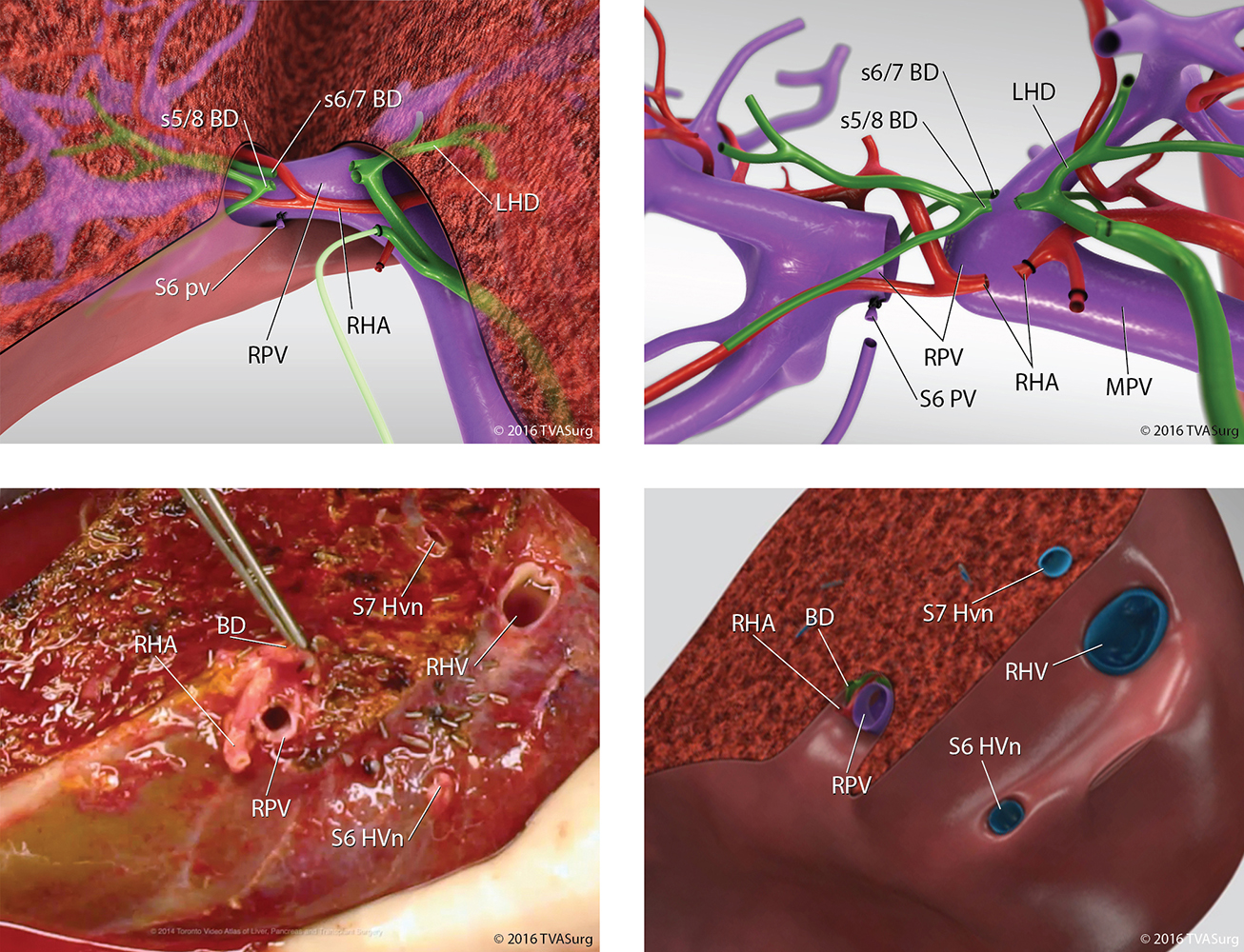
Figure 6 from the publication: Vascular and biliary divisions (A) 3D model render of porta hepatis dissection and division of bile ducts; (B) 3D model render of porta hepatis division locations; (C) photo of divided structures of extracted donor liver graft; (D) 3D model render of divided structures of extracted donor liver graft.
