Resection of Type II Choledochal Cyst
With Choledochoscopy
00:14 SURGICAL PLANNING
02:22 PORTAL DISSECTION
04:07 CHOLECYSTECTOMY & CHOLEDOCHOSCOPY
05:19 CYST EXCISION
Case description
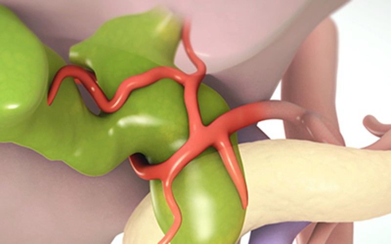
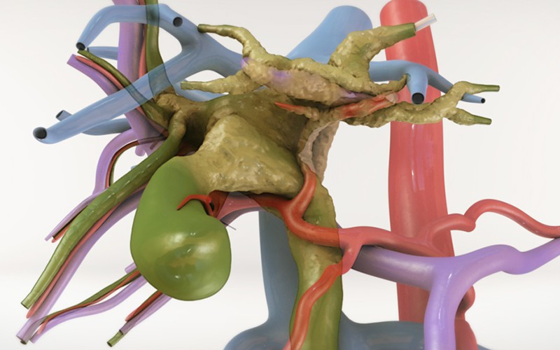
- The patient was a 39-year-old female and MRI findings suggested she had type II choledochal cyst, which is an outpouching of the extra hepatic bile duct. The patient also presented with choledocholithiasis.
- The surgical plan was to excise the section of the common hepatic duct above and common bile duct below the cyst. Any residual stones in the common bile duct was flushed with saline. Choledochoscopy was performed to ensure no residual stones in the common bile duct.
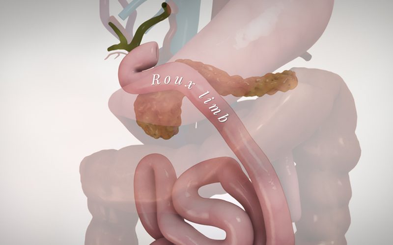
- After the cyst was excised, a Roux-en-Y hepaticojejunostomy was performed to re-establish bile flow.
MRI
Click to turn annotations on/off
Pathology
Radiology diagnosis thought to be type II choledochal cyst but pathology findings showed otherwise. Sections of the multiloculated cystic lesion show proliferating mucin-secreting columnar epithelium with underlying compacted cellular (ovarian-like) stroma. Multiple sections examined show no evidence of invasive carcinoma or high grade dysplasia.


