CASE DESCRIPTION
  • The flap design is marked on the forearm, with a 'S' shape extending from the antecubital fossa to the proximal margin.
  • The PL and FCR tendons are dissected at the ulnar border.
  • The cephalic vein is dissected from the proximal margin of the flap to the antecubital fossa.
  • At the radial border, cephalic vein branches that drain the skin flap are preserved, while the cephalic vein is divided.
  • The radial nerve is identified and preserved.
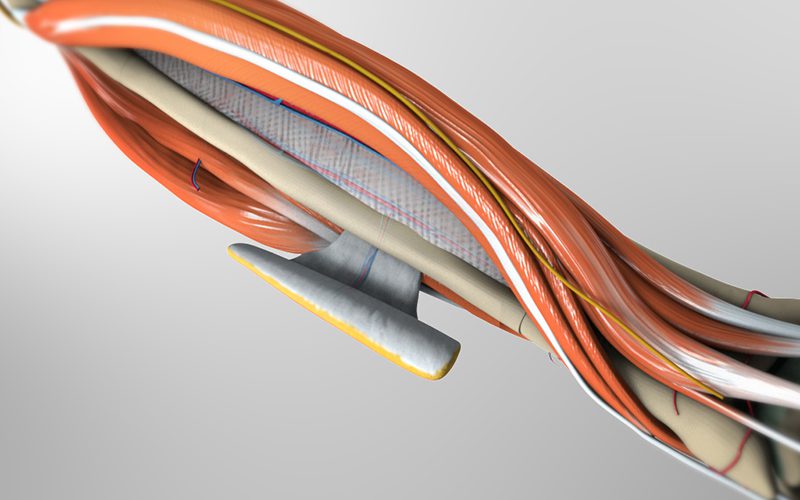
  • The BR tendon is identified, and the fascia between the BR muscle and FCR muscle is divided to reveal the radial artery pedicle. The radial artery pedicle is dissected towards the antecubital fossa.
  • At the distal margin of the flap, the radial artery pedicle is divided.
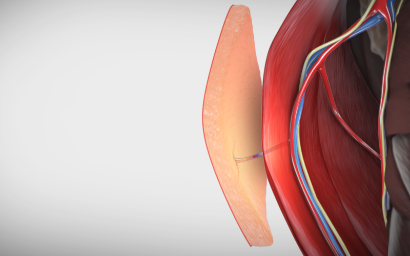
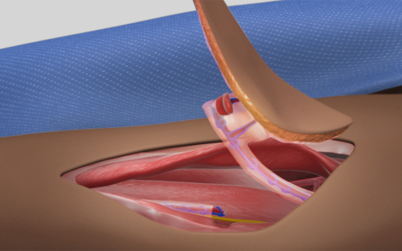
  • The skin flap is raised, following a dissection plane above the deep fascia. This ensures that perforator branches from the pedicle supplying the skin flap are preserved.
  • The pedicle is mobilized by dividing the inferior and lateral perforators.
  • The pedicle and cephalic vein is divided proximally near the antecubital fossa, and the free flap is removed for use in reconstruction.
PEARLS

Preoperative Assessment
  • Allen’s test should be performed to confirm ulnar arterial supply and radial artery patency.
  • Allen’s test using a pulse oximeter checking the waveform and oxygen saturation of the fingers, particularly, thumb and index finger can be used to confirm the visual Allen’s test. Alternatively, doppler of the arterial supply to the thumb can be assessed with a hand-held doppler with and without ulnar compression.
  • In patients with history of prior radial arterial cannulation (i.e. arterial line or catheterization for interventional radiology procedures) a reverse Allen’s test with compression of radial will assess occlusion of the radial artery. In cases where radial artery occlusion is suspected, doppler can be used to assess proximal flow and the flap can be redesigned and harvested more proximally1.
Flap Design
  • Incision is planned proximal to the flexor crease of wrist in the distal third of the forearm in order to capture distal radial artery perforators. Often will plan the distal aspect of the flap 2 to 3 cm proximal to the crease rather than at the crease itself. There are two dominant clusters of perforators from the radial artery, which occur in the distal third and proximal fifth of the forearm2.
  • We tend not to place the distal aspect of the flap 2cm or more from the flexor crease, but also note,based on surface landmarks when possible, where there are venous communicators from the cephalic vein to the RFFF.
  • There are relatively few perforators in the mid-forearm corresponding to the area of overlap between brachioradialis and flexor carpi radialis.
Flap Elevation
  • Special attention should be paid to the deep neurovascular structures of the anterior compartment of the forearm including the ulnar nerve, ulnar artery, and median nerve
  • The ulnar nerve generally is not at risk when the flap is placed proximal to the flexor crease.
  • Rarely, the ulnar artery can arise above the elbow and travel superficial to the flexor muscles of the forearm3.
  • In the distal forearm the ulnar artery can be superficial. We preserve the fascia overlying the muscles and tendons ulnar to the flexor carpi radialis tendon.
  • The median nerve travels deep to flexor digitorum superficialis for most of its course, but distally it courses lateral to the muscle and takes a more superficial course4. A persistent median artery can travel with the median nerve and provide its blood supply and blood supply to the superficial palmar arch.
Pedicle Dissection
  • When the cephalic vein is incorporated into the pedicle it is important to capture veins that communicate from the flap to the cephalic vein. If these are not present the cephalic vein can still be used but should be divided prior to dividing the main pedicle to assess flow. If flow is absent our practice is to not use the vein for anastaomsis.
  • The radial artery is accompanied by two draining venae comitantes. Proximally, these may join into a single vein which is often of larger caliber.
  • Often there is also a communication between the venae comitantes with the cephalic vein.
  • Surgeon preference dictates whether to use one draining vein versus performing separate anastomosis for the cephalic and venae comitantes
  • The radial recurrent artery limits the extent of proximal dissection of the radial artery. It is the first branch of the radial artery in the proximal forearm travels medial to the elbow and forms an anastomosis with the radial collateral artery.
Modifications
  • Sensate flaps may be harvested by incorporating sensory nerves of the forearm into the flap elevation. The lateral antebrachial cutaneous nerve is typically used.
  • Palmaris tendon can be incorporated into the flap harvest as well. Presence of the tendon can be assessed using Schaeffer’s test. The patient opposes their thumb fifth digit then flexes their wrist. The wrist is examined for presence of palmaris longus tendon medial to the flexor carpi radialis tendon. The tendon is absent in approximately 12% of the general population, with high variability according to ethnicity. Higher absence rates (15-30%) are observed in Nigerian5, Turkish6, Indian7, and American Caucasian populations8. Lower absence rates (2-5%) have been observed in Chinese9 and African-American populations8.
  • To obtain additional bulk a proximal extension to include adipofascial tissue over the mid-forearm can be performed. In these harvests care must be taken when dissecting along the proximal border of the skin flap to avoid disruption of the blood supply to this extension.
  • A portion of the radius bone can be harvested by preserving the inferior perforators during flap elevation over the retinaculum. Thickness of bone harvest should be limited to 50% of the diameter. A keel or boat-shaped osteotomy is typically employed and prophylactic internal fixation is performed to prevent fracture10.
Closure
  • When closing the donor site with a split-thickness skin graft avoid pie-crusting over the tendon.
  • It is best to cover the radial nerve prior to skin graft by advancing lateral forearm skin. Often the donor site  defect can be reduced in size through advancement of skin or purse string suture.
  • A bolster is applied over the skin graft for one week. Use of wound VAC has been reported, but caution should be employed to prevent compression of the ulnar artery and bloodflow to the hand11.
  • Where possible closure may also be accomplished using the hatchet modification12 or ulnar artery based perforator closure, such as the keystone flap13,14. Care must be taken to make sure any closure does not compromise ulnar flow to the hand and fingers.
References
  1. Foreman A, de Almeida JR, Gilbert R, Golstein DP. The Allen’s test: revisiting the importance of bidirectional testing to determine candidacy and design of radial forearm free flap harvest in the era of trans radial endovascular access procedures. J Otolaryngol Head Neck Surg. 2015 Nov 4;44:47.
  2. Saint-Cyr M, Mujadzic M, Wong C, Hatef D, Lajoie AS, Rohrich RJ. The Radial Artery Pedicle Perforator Flap: Vascular Analysis and Clinical Implications. Plast Reconstr Surg. 2010 May;125(5):1469-78.
  3. Fatah MF, Nancarrow JD, Murray DS. Raising the radial artery forearm flap: the superficial ulnar artery “trap”. Br J Plast Surg. 1985 Jul;38(3):394-5.
  4. Drake, RL, Vogl W, Mitchell AWM,and Gray H. Gray’s Anatomy for Students. Philadelphia. Elsevier/Churchill Livingstone, 2005. Print.
  5. Oluyemi KA, Adesanya OA, Odion BI, Ukwenya VO. Incidence of Palmaris Longus Muscle Absence in Nigerian Population. Int J Morphol 2008;26(2):305-8.
  6. Kose O, Adanir O, Cirpar M, Kurklu M, Kourcu M. The prevalence of absence of the palmaris longus: a study in Turkish population. Arch Orthop Trauma Surg 2009 May;129(5):609-11.
  7. Aggarwal P. Absence of the palmaris longus tendon in Indian population. Indian J Orthop. 2010 Apr;44(2):212-5.
  8. Soltani AM, Peric M, Francis CS, Nguyen TTJ, Chan LS et al. The Variation in the absence of the palmaris longus in a multiethnic population of the United States: an epidemiological study. Plast Surg Int 2012:2012:282959.
  9. Sebastin SJ, Lim AYT, Wong H-B. Clinical Assessment of Absence of the Palmaris Longus and its Association With Other Anatomical Anomalies – A Chinese Population Study. Ann Acad Med Singapore 2006;35:249-53.
  10. Shnayder Y, Tsue TT, Toby EB, Werle AH, Girod DA. Safe Osteocutaneous Radial Forearm Flap Harvest with Prophylactic Internal Fixation. Craniomaxillofac Trauma Reconstr. 2011 Sep;4(3):129-36.
  11. Andrews BT, Smith RB, Chang KE, Scharpf J, Goldstein DP, Funk GF. Management of the radial forearm free flap donor site with the vacuum-assisted closure (VAC) system. Laryngoscope 2006 Oct;116(10):1918-22.
  12. Lane JC, Swan MC, Cassell OCS. Closure of the radial forearm donor site using a local hatchet flap: analysis of 45 consecutive cases. Ann Plast Surg. 2013 Mar;70(3):308-12.
  13. Behan FC. The Keystone Design Perforator Island Flap in reconstructive surgery. ANZ J Surg. 2003 May;73(3):112-20.
  14. Potet P, De Bonnecaze G, Chabrillac E, Dupret-Bories A, Vergez S, Chaput B. Closure of radial forearm free flap donor site: A comparative study between keystone flap and skin graft. Head Neck 2020 Feb;42(2):217-223.

SHARE THIS: