Microvascular hepatic artery reconstruction
Vascular reconstruction technique for pediatric liver transplant
00:06 TECHNIQUE OVERVIEW
01:37 Exposure
03:10 Anterior wall
05:32 Posterior wall
Technique description
- During living donor liver transplantation, microsurgical techniques in arterial reconstruction greatly reduce the incidence of hepatic artery thrombosis.
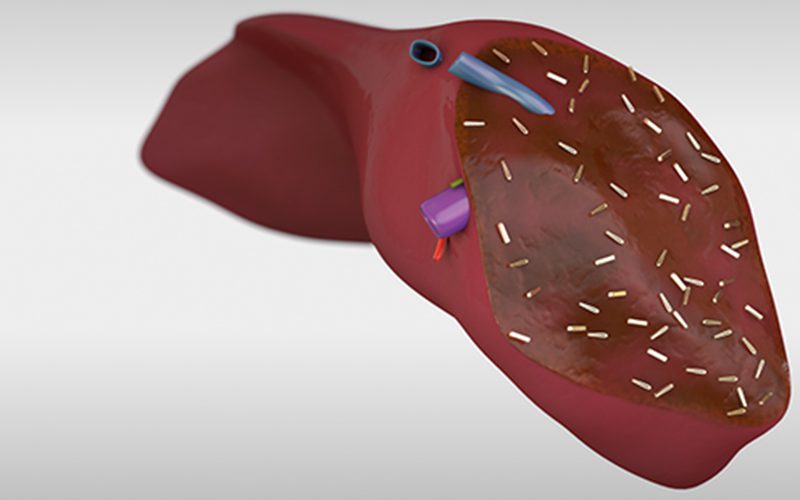
- This video will demonstrate the technique for an end-to-end arterial anastomosis for a living donor left lateral segment liver graft implantation into a pediatric recipient.
Key points
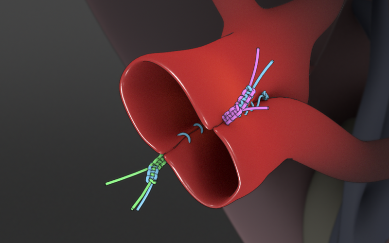
- The anastomosis will be completed with 8 interrupted stitches of 9-0 nylon sutue
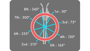
- In pediatric recipients of adult left lateral segment grafts, the recipient right hepatic artery is often a better choice for arterial revascularization of the graft since it is longer, larger, and has better inflow than the recipient left hepatic artery.
- As with portal vein reconstruction, redundancy in the arterial anastomosis is permitted to prevent kinking and twisting when the graft is oriented in its final resting position.
- Prior to suturing, the recipient hepatic artery will be gently dilated to reduce spasm.
- When tying, gentle lateral tension should applied to the sutures in a direction perpendicular to the axis of the vessels; the amount of tension should be equal on both ends.
- Intermittent counter-tension should be applied in the direction of the axis of the vessels; this helps distribute tension evenly along the suture.
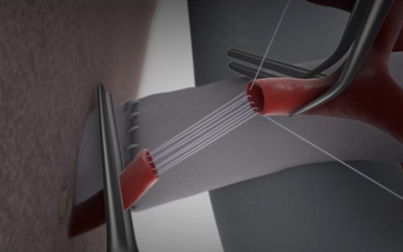
- An alternative method for hepatic artery reconstruction, when deemed appropriate, is the parachute technique.
References
- Reducing Pediatric Liver Transplant Complications: A Potential Roadmap for Transplant Quality Improvement Initiatives Within North America
- "Hepatic Artery Reconstruction in Living Donor Liver Transplantation From the Microsurgeon’s Point of View" – Furuta et al.
- Arterial end-to-end anastomosis of the rat carotid artery with 8 interrupted sutures (according to Yonekawa)

