
Laparoscopic right hepatectomy
With ultrasonic dissection and Pringle maneuver
00:12 Surgical Plan
02:25 Patient position and port placement
03:34 Mobilization and cholecystectomy
05:46 Pringle maneuver setup
06:56 Portal dissection
Case Description
- The patient is 76 years old, diagnosed with colon cancer with liver metastases.
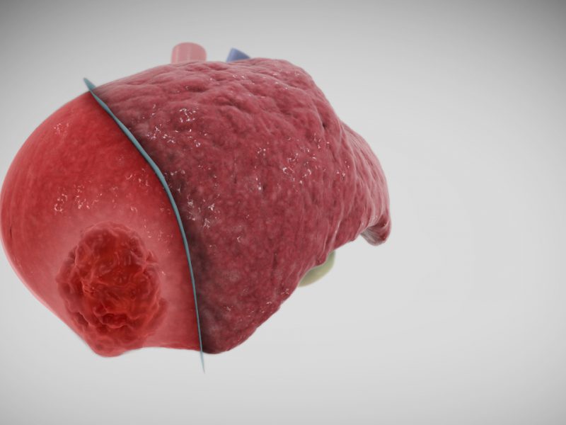
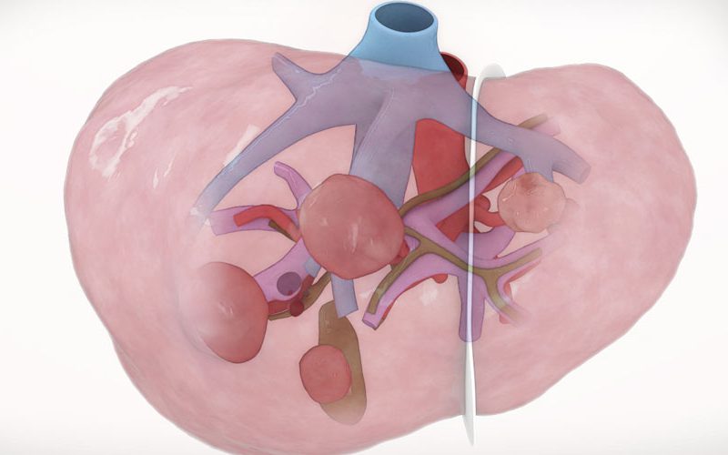
- The tumor involved liver segments 5 to 8, and was pressing against the underside of the liver.
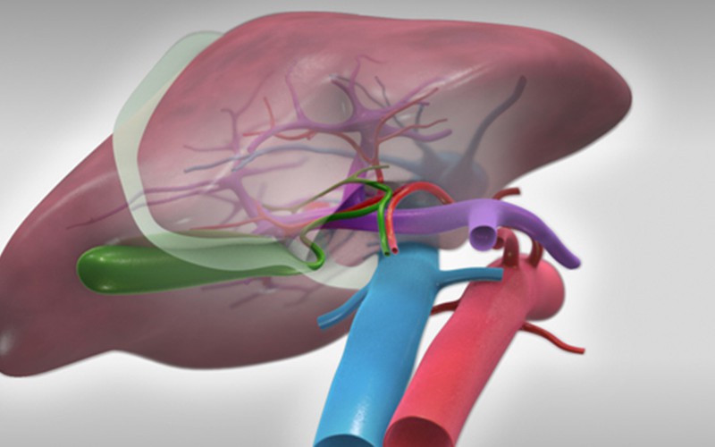
- A laparoscopic right hepatectomy was planned, using an ultrasound dissector as well as the Pringle maneuver to occlude portal and hepatic artery inflow.
- To optimize visualization during mobilization of the right lobe liver, the patient is positioned in a reverse-Trendelenburg position with the right side up, with the aid of an inflated saline bag under the right flank.
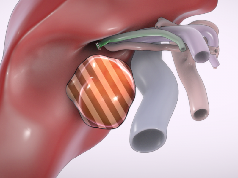
- During portal dissection, traction is applied by pulling the gallbladder cephalad, while counter-traction is applied by pulling the umbilical tape in the Pringle maneuver caudad. Additional exposure can be obtained by pulling on the cystic duct stump, previously ligated with vascular clips.
- With the laparoscopic caudal view, the caudate lobe can be easily accessed. Transecting the caudate lobe in this view allows for easy mobilization and division of hilar structures such as the biliary plate.
CT scans (venous)
Click to turn annotations on/off
CT reconstruction model
This is the raw CT reconstruction model set made with Intrasense Myrian for this case. We use automated reconstructions for the base level models, and then retopologize using modelling software. This model set represents the first stage in our 3D modelling workflow.
Thanks
Very nice.