Laparoscopic Liver Donor Right Hepatectomy
with the use of indocyanine green (ICG) fluorescence
00:14 Introduction and surgical plan
02:45 Patient position
03:41 Exposure and mobilization
06:49 Portal dissection
07:25 Parenchymal dissection
Case Description
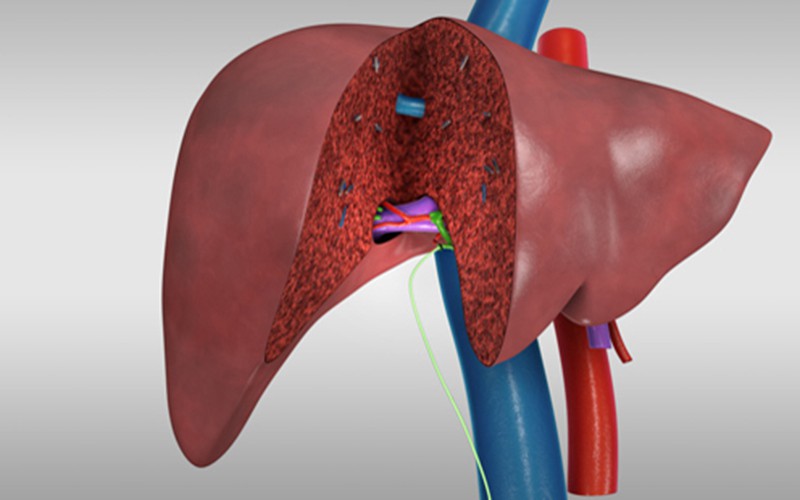
- The patient is a 34-year-old female scheduled to undergo a living donor laparoscopic right hepatectomy for an adult recipient.
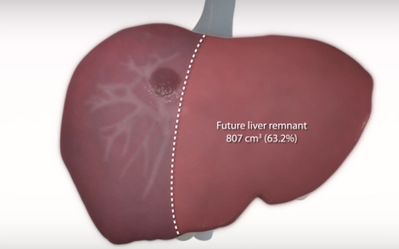
- Preoperative planning determined that adequate remnant volume would require leaving the middle hepatic vein in the donor, resulting in 33% future liver remnant.
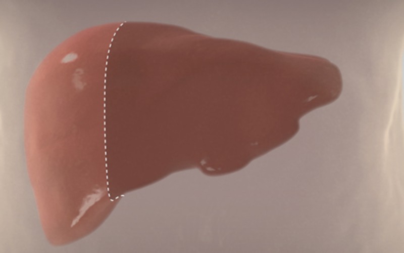
- The planned transection plane extends from the right-middle hepatic groove through the gallbladder fossa.
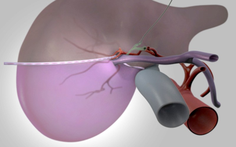
- To confirm the line of transection, ICG fluorescence is used to visualize the margin of the right hepatic lobe.
- ICG fluorescence is also used to guide the right hepatic duct division.
- The patient is placed in a right semi-lateral modified lithotomy position.
- A double-lumen endotracheal tube is used to independently ventilate the left lung.
- A right lumbar bean bag is used to elevate the right lobe to aid in the mobilization of the right lobe.
CT scans (venous phase)
Click to turn annotations on/off
CT scans (arterial phase)
Click to turn annotations on/off
MR Reconstruction Model


