In Vivo Lung Perfusion
00:12 Surgical Plan
04:00 Surgical exposure
04:47 Hilar dissection
05:35 In Vivo Lung Perfusion
06:11 Cannulation
07:05 Lung perfusion setup
07:28 Perfusion system
08:12 Washout
08:49 Closure
Case Description
- The patient was a 20-year old woman, diagnosed with multiple metastatic lesions from sarcoma in both lungs.
- In Vivo Lung Perfusion (IVLP) will be used to isolate the left lung and deliver a high dose of chemotherapy without systemic exposure.
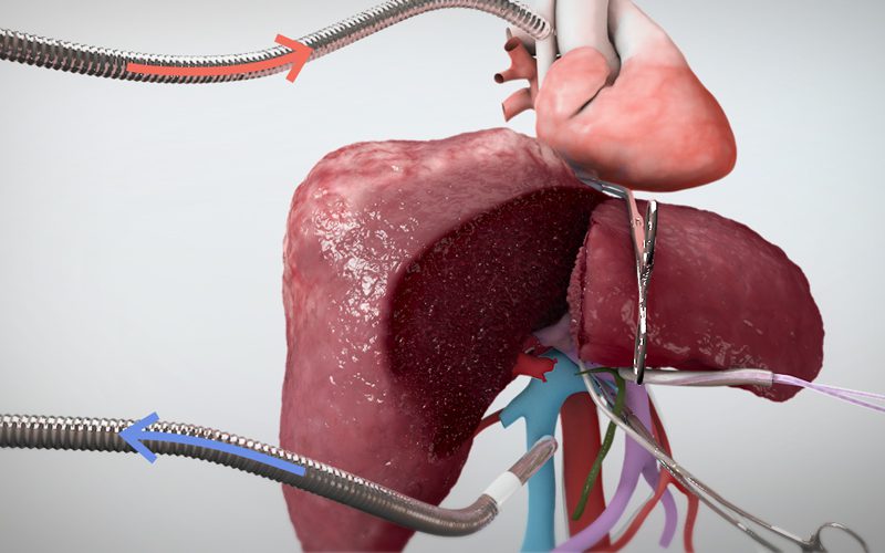
- The left lung is deflated to expose the hilum and the left bronchus, pulmonary artery (PA) and pulmonary veins (LUPV and LLPV) are exposed.
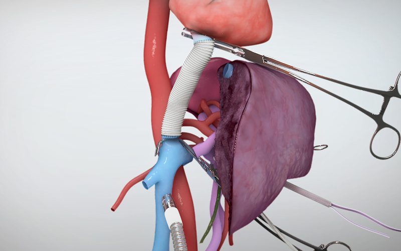
- After administration of heparin, the LPA, LUPV and LLPV are cannulated.
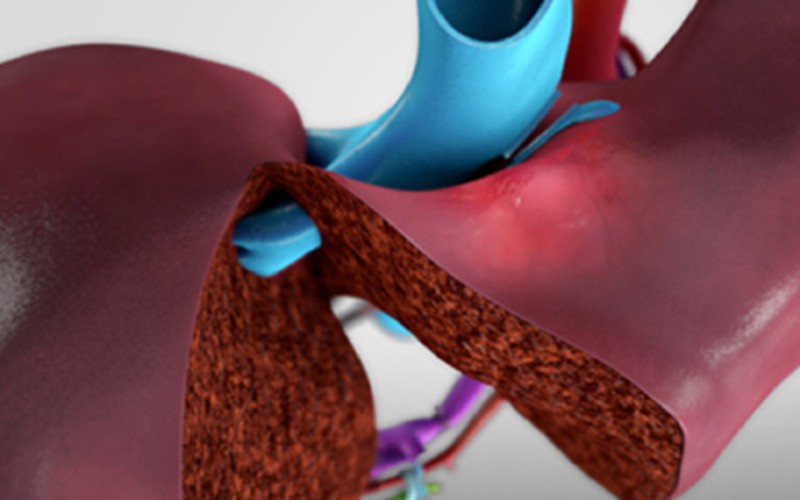
- The LPA is ligated with a tourniquet clamp, and the LPVs are interrupted with a vascular clamp beyond the vein cannulation.
- The left lung is re-ventilated for the perfusion period.
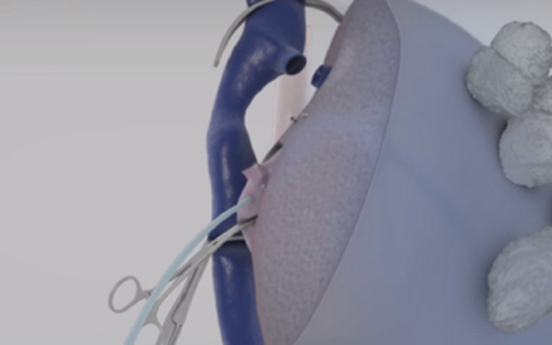
- Blood leaving the left lung is diverted through the cannulas to a hard shell reservoir, where chemotherapy is administered, forming perfusate.
- The perfusate is then directed to a centrifugal pump, which drives the flow to a membrane gas exchanger, which is connected to a heater/cooler.
- At the membrane gas exchanger, a gas combination is added to deoxygenate the perfusate and provide CO2 for the inflow.
- The perfusate passes through a leukocyte filer and returns to the LPA through the cannula.
- After 3 hours of IVLP, the circuit is disconnected from the chemo, and connected to a bag of Perfadex to flush the lung in one pass.
- The pulmonary artery tourniquet is opened gradually to de-air the lungs, the cannulas are removed and venotomy sites repaired.
CT scans (axial)
Click to turn annotations on/off
CT scans (coronal)
Click to turn annotations on/off
Animation models (retopologized)
Shown here are the models used in the animation for this video, based on automatic 3D reconstructions of the pre-op scans.