
Apart from our bread-and-butter educational surgical videos, the TVASurg team also collaborates with our staff surgeons, scientists and surgical fellows in academic publications, by creating medical illustrations to accompany their articles.
Below are a selection of pieces that have been published in journals such as Nature Communications, Liver Transplantation, Transplantation, and the British Journal of Surgery.
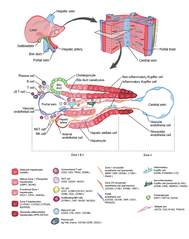
Journal published in: Nature Communications
Accompanying article: https://doi.org/10.1038/s41467-018-06318-7
Medical illustrator: Joy Qu
This illustration depicts a map of the human liver at single-cell resolution. The goal was to capture the intricacies of the liver at three distinct levels – organ, tissue, and cell – with a primary focus on the cellular level. Directional cues in the form of arrows lead the viewer between the three different biological levels.
The main challenge was to maintain cohesiveness while incorporating colour variation throughout the illustration for different cell populations. Conventional colours were used for the hepatic artery, vein, and bile ducts, while a more muted colour scheme was chosen for liver infiltrating lymphocytes. Furthermore, variations in colour value are used to distinguish clusters of gene expression within the same cell type (ex. hepatocytes).
Journal published in: Liver Transplantation (cover image)
Accompanying article: https://doi.org/10.1002/lt.23986
Medical illustrator: Paul Kelly
This illustration was created to explain the key components of the Ex Vivo Liver Perfusion system developed by the Liver Transplantation research team at Toronto General Hospital. Dr. Markus Selzner and Ian McGilvray are two of the lead surgeons involved in this research and acted as content experts on this illustration.
The visualization approach was to indicate key components of the apparatus using outlines, placing them visually in relation to one another so that the circulation of the system took precedence, and the surface details of these mechanical components did not pull the viewer’s attention away from the pathway flow in the depiction. Important phases of the system’s circulation pathways were colour-coded to show their analogous nature to liver blood circulation, and text details specific to each circulation phase were overlaid on the pathways to conserve space. The liver was rendered using 3D software to give it visual prominence and provide a focal point for the piece. A posterior view was utilized to provide maximum access to vessels involved, and for the sake of accuracy, as liver grafts within the Ex Vivo Liver Perfusion system are usually laid flat in a facedown manner.
Journal published with: Transplantation (cover image)
Accompanying journal: June 2020 – Volume 104 – Issue 6
Medical illustrator: Albert Fung
The editorial image was created for an issue of the Transplantation journal which contained a series of consensus papers highlighting transplant oncology, which utilizes liver transplantation as a treatment option for patients.
Consideration was taken to highlight the specific nature of the field of transplant oncology, which resulted in a highly contrasted depiction between a cirrhotic, tumor-ridden liver (left), and a healthy liver graft (right). In the center background is the standard inflow/outflow occlusion setup of the hepatic veins, hepatic arteries and portal veins during a liver transplant procedure, providing context between the specimen and graft.
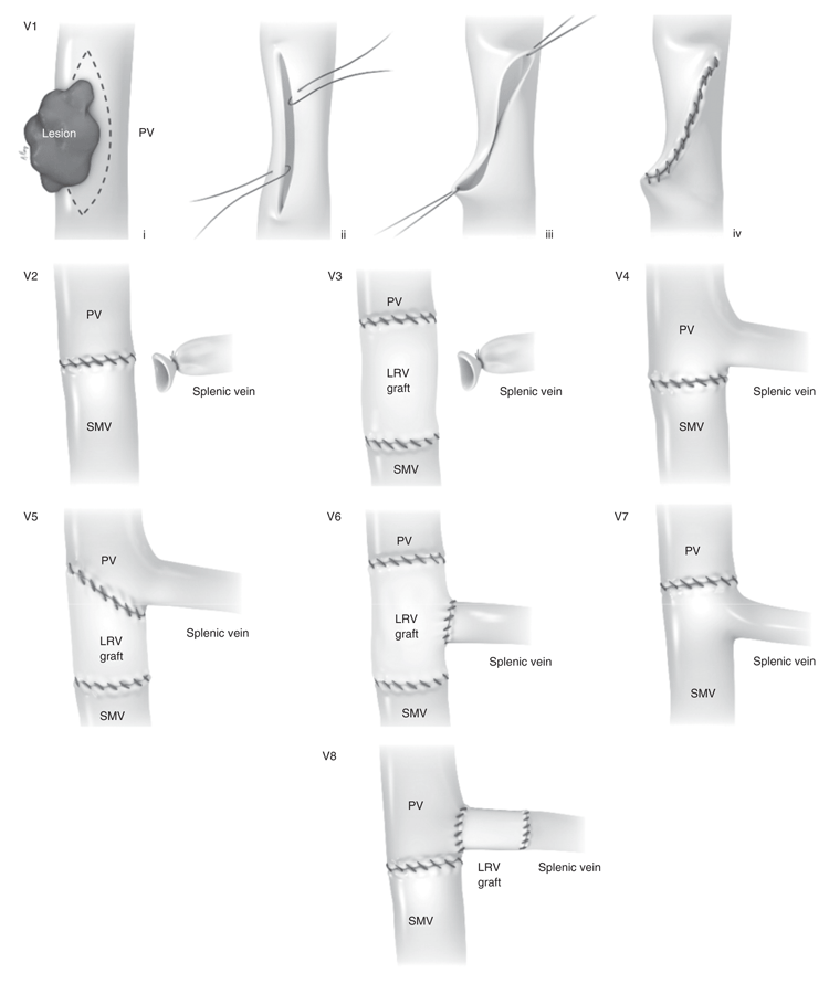
Journal published in: British Journal of Surgery
Accompanying article: https://doi.org/10.1002/bjs.9222
Medical illustrator: Albert Fung
This series of images illustrates the variety of different options to reconstruct the portal vein during a Whipple procedure, where a resection of the vein may be required to achieve negative margins.
Depicting the majority of the options (V2 – V8) was relatively straight-forward, where displaying the end-result of the vein anastomoses, along with appropriate labels, was sufficient to understand the approach. As a comparison, option V1 required a sequential illustration to convey the steps taken in removing the tumor, and closing the defect in the vein wall.
As colleagues of outstanding physicians and scientists, we’re always excited to convey their work into aesthetic, visual pieces, with hopes to help them disseminate their messages in their academic careers.
The TVASurg team