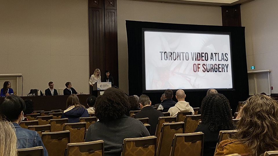
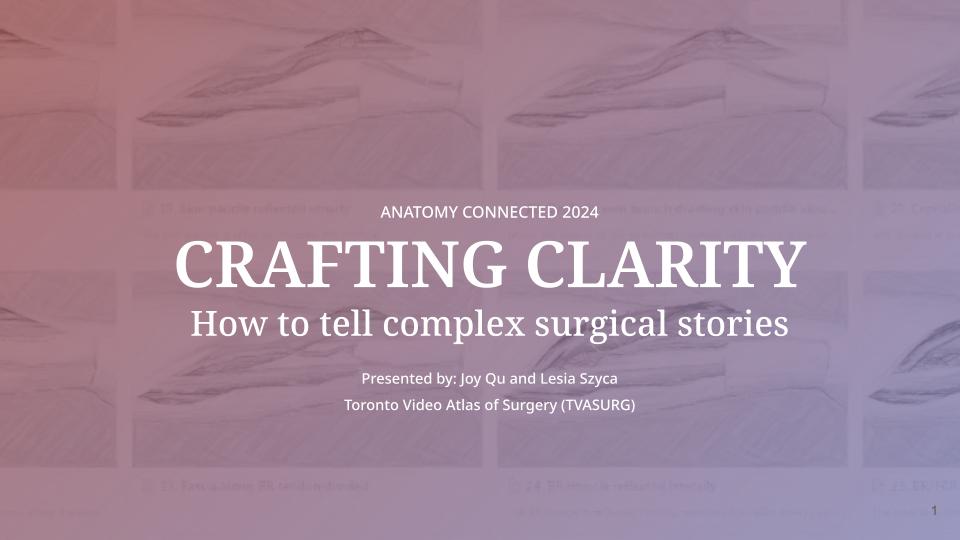
We recently attended the American Association of Anatomy's2024 Anatomy Connected conference held this year in Toronto. TVASurg team members Joy Qu and Lesia Szyca presented with other medical illustration professionals at the Association of Medical Illustrators-sponsored group session "Design Thinking and Visual Communication through the Lens of the Medical Illustrator". TVASurg team member Paul Kelly, and host of the Medical Illustration Podcast was also in attendance and has compiled a podcast about the event:
Day 1: Friday, 22nd of March 2024
On the evening of Friday the 22nd of March 2024, the event kicked off with a plenary session by Dr Kurt Schwenk, from the Department of Ecology & Evolutionary Biology at University of Connecticut, who presented on using micro videography to study precise movements of lizards in capturing prey. This is a good place to point out that the work of anatomists can span a wide range of species, not just humans. He worked as a zookeeper in Brooklyn NY while pursuing his PhD. His focus more recently has been on the lizard tongue. This was high speed anatomy in motion. Really cool and presented with lots of histological images from the lab and high-speed video clips, so you could see these unique tongues, composed of pure muscle tissue taking on dimensional changes with conservation of volume throughout complex motions for prey capture. This line of study looked into the biological adhesive qualities and unique structural qualities such as the hexagonal shape of tongue papillae allowing for maximum packing. We got to see the Chameleon tongue “bedewed with viscous secretion” changing shape before it even contacts its prey. Pretty wild!
Day 2: Saturday, 23rd of March 2024
Before attending the conference I noted in the program that AI was a feature in many presentations. These began on Saturday morning with a panel discussion–a set of presentations from Patrick Pennfather, assistant professor for Theatre and Film at the University of British Colombia. He was quite enthusiastic about the prospects for AI, and shared some of his personal journey which included early investigations into fish movement patterns. Interestingly enough I’ve heard people talk about that before being a useful application for these kinds of tools. His motivations were to investigate the process of developing technology that solves a human problem. So we need to identify problems to solve, and then we can identify generative AI as a potential solution if we understand the challenges which include the complexity of science, the potential for misinformation bias, of course, data privileges, hallucinations of the AI, and public skepticism. He concluded with a recommended actionable item, which was to spend more time and focus on data organization and metadata labeling, which I thought was interesting because metadata is something leaders in our field such as Tonya Hines have been encouraging for quite a while now, so I definitely see how that would translate into our field.
Next up we had Michael Hickman, one of my former podcast guests from the Barrow Neurological Institute in Arizona, presenting on “Navigating AI in Medical Illustration, Some Considerations.” I appreciate the candid nature of Michael’s talk–he started right off at the get-go talking about the concern of undercutting the market in pricing. We are now looking at a situation where low costs and high volumes will become the norm. Michael encouraged more partnerships with medical illustrators, because we as medical illustrators have been able to navigate massive sweeping changes in our industry that were analogous to current AI concerns. Current and previous generations of medical illustrators went through a revolution with medical photography, this was something another one of my former guests Mesa Schumacher commented on where we went through a shift where we were no longer responsible or needed to document things, because we had come up with technology that could document things faster and more efficiently (essentially photography and video) so our role shifted more into interpretation, simplification, distilling information and depicting what cannot be seen. The next big revolution in medical illustration was the move to digital–where traditional media now had to take a backseat to tools that allowed for endless undo actions, filters, colour corrections, layers, and incredible degrees of control, and of course repurposing of assets. Most recently, the field of medical illustration has integrated 3D models and animation as a new forefront of our technological tool kit.
So as we continue to adjust to changes in the tools used for image creation, we have learned to lean in more to one of our super powers, which is our ability to design and depict new subject matter, and things that have never been previously visualized. This is something that AI is still not really able to do very well, as Generative AI is based on essentially re-mixing already existing content that has been scraped from the Internet. So this segues into the ethics concerns of using AI. We do have some options. There are tools, such as Glaze which acts as a protective measure, adding a barely discernible filter over images, which will make them difficult to detect and categorize by AI scraping algorithms. Nightshade is a more offensive tool, impairing AI generative models’ ability to create images by essentially “poisoning'' a dataset. From their website:
“Nightshade's goal is not to break models, but to increase the cost of training on unlicensed data, such that licensing images from their creators becomes a viable alternative.”
Ultimately, I do think the option most of us will opt for is the so-called “ethical” AI models such as Adobe Firefly, which are paid services, but are designed for reimbursing artists for their contributions to a dataset. It’s also worth noting that as biomedical communicators and scientific visualizers, we have an awareness and familiarity with the medical terminology that is used in our work. This may include some words that are restricted from use in publicly open, generative, art models, such as Midjourney, which do have a degree of censorship over certain terms. I thought Michael did a great job of laying all this out for us to think about.
The next speaker on this panel was Sean Seung Hyun Jeon, Product Manager at UBC HIVE speaking on AI as a tool in anatomy education. He began by pointing out that while large language models that have been fed massive amounts of data, have become a cause of concern–but if we take a step back and think about when Google first came out, we also had a fear of what that was going to mean to our jobs, and humans ability to keep pace. What we need to ask though is “why?” Why are we using these tools? Why are we concerned? Why are they the best option? Sean shared a helpful acronym for interfacing with AI tools: THINK. Is what this tool telling me True? Is it Helpful? Is it Inspiring? Is it Necessary? And is it Kind? He also shared an important observation I think we should all be keeping in mind with AI generative tools which is a Western bias in a lot of what we’re seeing in both the images and in the text generated. These tools having been developed in the United States, I think there’s of course going to be a bias there and we need to be aware of this. He closed by making the point that ultimately the teacher is more important than the tool or the tech and I would definitely agree with them there.
The next presenter was Bailey Lo from the Biomedical Visualization communications program at UBC, who is also a fellow podcaster I might add, speaking about generative AI in science communication. She was speaking from a learning experience designer perspective, making connections between pedagogy and technology. She started off by saying that our problems statements need to come before our solutions, and I couldn't agree more. I was also happy to hear this theme repeated throughout the conference. This has been my sentiment as well as it’s great when technology comes out and it has all these new fancy capabilities but if it’s not solving a specific problem, then it’s not necessarily useful to us.
Bailey also echoed Sean’s observations that there is a lack of diverse inputs in the AI and equity is never addressed as a computational problem. Some suggested use cases for AI include building rough drafts, coding, cost in time estimates for work. I think these are useful applications, and are similar to the way in which I have been using tools like ChatGPT recently. Although I hadn’t thought to use it for costing time estimates— that’s actually a good idea. I think I’m gonna try that.
But at the end of the day we need to think about methods and strategies to combat and regulate misinformation. We’re already in a situation where we’re probably seeing AI generated images fed back into AI art generation models which is going to compound inaccuracies. We need to emphasize human compassion and let’s choose skeptical curiosity over skeptical criticism.
Design Thinking: Visual Communication Through the Lens of the Medical Illustrator
The next session was “Design Thinking: Visual Communication Through the Lens of the Medical Illustrator.” This was the AMI sponsored session spearheaded by Jill Gregory at ICANN School of Medicine in New York City. The session began with Chris Smith, describing how “The Medical Illustrator’s Toolkit Drives New Approaches in Anatomical Research”. Chris is a high-level illustrator from the Johns Hopkins program, now a postdoctoral fellow at the Division of Anthropology, American Museum of Natural History, in New York, who has recently been digging deep into research on the evolution of the inner ear. Chris began by giving a bit of a background on medical illustration as a field. He talked about our role as visual storytelling, and how we create what cannot be seen or what has never never been seen and he also described some of our transformative modalities, which include thinking in 3D, modular approach, and shifting perspective. This really speaks to strong fundamentals in all aspects of visual creation. He shared a tip from one of his professors, which I thought was particularly useful for everyone in medical illustration to consider: “Can someone sculpt what you drew?”
Chris then went on to speak more about the research he’s been doing in particular regarding the vestibule and semicircular canals of the inner ear apparatus. One takeaway that I got was, I don’t think I’d heard of this structure before—the membrana limitans—a membrane lying just inferior to the utricle, providing support and acting as a structural boundary between portions of the vestibule. This structure being involved with balance and orientation. I was blown away by Chris’s ability not only to perceive some of these minute structures from scan slices but also to find clever angles to pose this anatomy to depict it for a viewer. Chris would go on to present again later at the conference, on Monday, on "Functional and Spatial Constraints in the Mammalian Vestibular System".


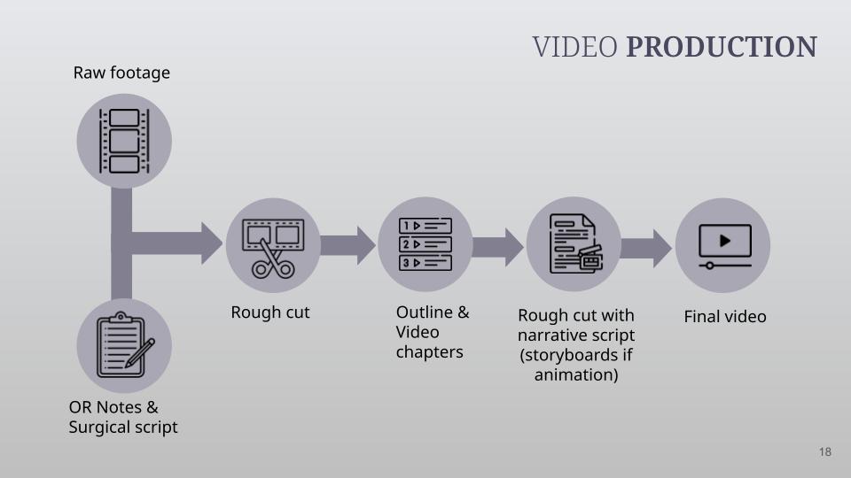
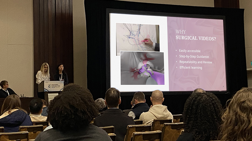
Next up were my colleagues from TVASurg, Joy Qu & Lesia Szyca, presenting on our work at the Toronto Video Atlas of Surgery in “Crafting Clarity: How to Tell Complex Surgical Stories.” I had the privilege to see this talk come together behind the scenes and I was equally impressed by the production and delivery. Joy and Lesia spoke about the benefits of the video medium for our subject matter, understanding our target audience, and the role of surgeons in our production process. They went into the art of surgical storytelling discussing how we enhance shots with video editing and compositing techniques, add clarity with visual cues, and enhance our content with 3D animation.
They did an amazing job and I was happy to see people line up to the mic at the Q&A after their talk. It’s always great to hear the feedback from people in the audience who share new insights and perspectives. I always walk away from these conversations with new ideas and questions and that’s what conferences are all about— they give you new considerations and curiosities about work you’ve been doing for extended lengths of time, so it really is a mental breath of fresh air.
Rounding out the session were Shay Saharan from University of Toronto and Naomi Robson from Bridgeable, presenting: “Beyond the Books: Exploring the Potential of Design, Interactivity and Learner-Centered Thinking in Anatomical Education”. This was a bit of a different approach in applying the tools and techniques of medical illustrators for what I took to be user experience design. They gave an overview of the production process, which I always love to see these depictions, especially when they start to dissect that process. A big focus of this talk was exploring the users and their needs, and iteratively evaluating and questioning their own assumptions.
So the iterative design approach and revision phases here were not being driven by client preferences or fine-tuning of accuracy–but rather fine-tuning the delivery mechanism for maximum effectiveness. Shay and Naomi discussed asking different evaluation questions at different phases—this I think does mirror the experience of working with medical content experts and at different points in production you have to direct the focus on certain types of revisions because otherwise, you’re just gonna be overwhelmed or you’re going to be distracted, working on content that will later only require further revision or omission. Shay also shared a link for SciViz tips and resources which I’ll share.
A beautifully composed group session, touching on different aspects of medical illustration: 2D illustrations, 3D linear storytelling and interactive media experience design, all for different audiences. I was inspired and incredibly proud to count myself among this group.
AI in anatomy education
As mentioned, AI was a hot topic across the entire conference program. I was so impressed with the measured response to AI throughout this event—I would describe it as optimistic curiosity with cautious evaluation—which is very much the approach I myself have been seeking to take. I saw throughout the presentations and Q&A follow-ups that educators know their students will be using AI and they will have to keep pace, but they are also asking the right questions imo, such as “What is the problem we are using this to solve?” and “How can we instruct students to use these tools effectively, but not be reliant on them?”
Many of the attendees have been in the field of education for a long time and they’ve already lived through the explosion of the internet and rapid integration of technology. They’ve seen the advancements and they’ve heard a lot of sales pitches. Not every new gadget pans out, but you also can’t ignore the rising tides.
Some of the tips from educators on how best to adjust to AI use in education settings included increased transparency, deciphering of diversity, evaluating uncertainty and variation, educator awareness, encouraging AI- free time, and looking for ways to extend human capacity. I think in the future, we may see customized lesson plans for individual interests, and custom AI models specific to certain education institutions.
Drew Berry keynote
The Saturday talks concluded with the keynote presentation by Dr Drew Berry. Drew has been a fantastic contributor to biomolecular animation for many years. In this talk he revisited some of his greatest hits and shared some of the new projects he’s been working on, which included some pretty massive videos for Omnimax theaters. So he’s doing 8K video, 60 fps, projected onto dome screens, and stereoscopic as well. That’s incredible to imagine and the computing power to produce that is also staggering. He walked us through some of the production process and how they had found a Unity plugin that worked for projecting onto the dome-shaped screens. I think I wasn’t the only person to raise an eyebrow at that because in case you haven’t been keeping up with industry trends, Unity has fallen out of favour after they changed their policies, and the Unreal game engine has become preferable to a lot of gaming and interactive developers, but Dr. Berry explained that they started making these Omnimax movies before all of that transpired and their workflow still works so they’ve stuck with it. Drew also spoke about the importance of sound design in his animations—he has a good friend who’s been a collaborator with him for a long time and who had worked on horror movies, so he’s familiar with using music and sound effects to evoke strong emotions. Another thing in his presentation I thought was interesting was his focus on the young adult target population, specifically high school age kids. I think that’s awesome that he’s looking towards the future and making content to inspire the next generation of biological scientists.
Research posters
Speaking of the future of science, I was so impressed with all of the research posters. There was a massive space dedicated to these. I want to give a special shout out to all of those who were traveling to Canada from international destinations. I loved seeing all of the anatomical variations and I was impressed by some of the illustrations I saw. I also liked how the schedule of this conference really encouraged engagement with the poster presenters. A few of note, “Anatomical Variation of Hepatic Arteries: Insights from a 13-year Cadaver Study in India” by Dr Chitra Ramasamy, with whom I got to speak with for a few minutes.
“Subscapular Artery Variations and Military Personnel Considerations: A Case Report and Review” I believe I got to speak with Olivia Staser the first author on this one and if I recall correctly she had done the illustrations as well, which were very good. “Fibularis Unicus: A Singular Lateral Leg Muscle” was cool. It's basically fibularis longus and brevis combined. I think first author Alexis Chandler was standing by this poster and I got to speak with her about it briefly.
One poster I thought was super cool was a “Quadrifurcating Celiac Artery” by Nancy Adams. That was awesome! So. the Common Hepatic artery, Left Gastric Artery, Splenic Artery and Right Hepatic Artery were all branching from the celiac trunk. That was fascinating. You just don’t see that.
Props to Sarah Gluschitz from St. George’s University on their poster, study, and accompanying illustration, “The Menstrual Cycle: Comparing and contrasting the linear and circular model through the lens of the medical illustrator” this one really stood out. Beautiful design and visualizations.
How far would you go for the perfect picture?
I wanted to note that one of the sessions I found to be discussed quite a bit out in the hallways, which I did not attend, was entitled “How far would you go for the perfect picture?” This being about ethical considerations in anatomy education. One of the speakers was Dr Sabine Hildebrandt from Boston Children’s Hospital and Harvard University Medical school, speaking on “Anatomy in Nazi Germany and the Pernkopf Atlas: How the Past Shines a Light on the Present and Leads to Change in the Practice of a Publisher.” I have previously attended this lecture at U of T so I didn’t go to this talk, but it’s worth mentioning as I say because it was notable for many of the people who attended it, and if you are unfamiliar with the history of the Pernkopf Atlas, it’s definitely worth looking into. I’ll include a link to a previous lecture on the topic by Dr Hildebrandt.
Day 3: Sunday, 24th of March 2024
I should mention that a lot of focus of the talks at this event were on curriculum design–understandably so as this was an academic conference of anatomy educators. This manifested in several ways. A lot of the talks were focussed around learning activities, comparing learning outcomes and assessments. These educators are constantly asking themselves questions such as “What is the essential anatomical knowledge that needs to be taught?” and “What is the clinical application of anatomy?” I didn’t attend all of these presentations as they didn’t always pertain to me, but I wanted to point out that was a major focus as you’ll soon see.
The first set of talks I attended on Sunday morning was the educational session which began with a surgical skills training presentation. Dr Danielle Brewer-Deluce from McMaster University compared human and pig models and different embalming fluid types which I hadn’t heard of before but I guess when you’re running a university level course on dissection, you have to take these things into account. So they compared four different embalming fluids, looking at mechanical wear-and-tear testing and preservation of visual appearance.
There was a presentation from Dr Nathan Tullos from the University of Mississippi Medical Center comparing photogrammetry with the WIDAR app scanning method. This is an app that uses your cell phone camera. The motivation for this, of course, being decreased time for lab and lecture. The presenter had experience with manual methods of photogrammetry using Metashape and Blender. When comparing the results, they found the photo time to be roughly the same, but the WIDAR produced models much faster and also with smaller file sizes, which made it easier to embed these model files in PowerPoint, which I didn’t realize you could do, but that’s pretty cool I will have to try that out.
Video for anatomy education
Next Eduardo Muscogliati from Hull-York Medical School presented “A Framework for High-Quality Educational Anatomy Video Production.” He shared the YouTube Quality Assessment metrics comparing 2 videos on hip joint anatomy, one using a YouTube Quality Assessment checklist and the other without. Both videos were about 12 minutes in length shot with the same equipment, one presented by himself and the other presented by his twin brother, which made for a unique twist on ensuring an adequate control group. They found that post-test results were higher for students who had viewed the video with YTQA and that video also had higher youtube views over time. I think he did note that could be because youtube was more likely to recommend that video that was in line with their guidelines, not necessarily because it was better but the point stands. If you know the requirements and standards of the platform you’re using, it’s going to make an impact on viewership.
I’m going to move on a bit to some other sessions, my apologies for speakers I’m skipping over. I have to give credit to the AAA for including not just one but at least 2 sessions I saw that were focused on reporting and reflections on Failure. This was refreshing and I felt therapeutic. I think this was a great idea to include in a conference. To be honest I sometimes forget that imposter syndrome is prevalent in all walks of life, not just for us medical illustrators. I think for us it feels really prominent because you can so easily and readily compare your work with someone else’s but that’s also true for people publishing papers or teaching classes. One presenter I saw, Dr David Morton of The Noted Anatomist youtube channel, talked about comparing ourselves to other people and their accomplishments and having to keep that in check. I think social media can be a real trigger for this for many people. He recommended using those feelings of self-consciousness and imposter syndrome as indicators to seek out learning, observe what others are doing and take cues from them, and look for opportunities to collaborate. I like the quote he capped this session off with “Humility is not thinking less of yourself, but thinking less about yourself.” Words to live by.
Advanced 3D Imaging Modalities
For the Advanced 3D Imaging Modalities session there was one that stood out to me I wanted to mention, this was a talk by Dr Mei Zhen, professor of molecular genetics at the University of Toronto, on the neuronal development of C Elegans, widely considered to be an ideal species for research and she explains why. I actually didn’t know this but this tiny worm only has 300 neurons in its adult state! This makes it very reasonable to get a grasp on what each cell in this organism is doing through different stages of development. In her research, Dr Zhen showed how they were able to observe rewiring events converting axons to dendrites and dendrites to axons, meaning that neuronal signaling actually reverses direction over the course of time, she described the process as “a gradual transition of mixed identity with perfect spatial coordination” I thought that was incredible! I also thought she had a beautiful hero image for the start of her talk and several simple and effective animations showing 1-3 nerve cells. I can honestly say I’ve never been so excited and fascinated by C Elegans before!
Later on Sunday, I attended another Education Platform Session. Kayla Vieno presented “Approaches for, and Outcomes Of, Anatomical Variation in Gross Anatomy Courses Across Degree Programs: A Scoping Review”. Anatomical variation being commonly related to medical errors. An explicit approach to presenting these is preferable to implicit. Explicit in this context meaning a more structured educational framework with learning timelines and objectives. As was mentioned in a lot of these talks, contact hours for anatomy in medical education are declining so they have to balance a focus on anatomical variants within time constraints.
The next in this session was “Decoding DPT Anatomy Education Trends in the U.S. a Journey into Student Satisfaction, Innovative Teaching, and the Power of Cadaver-Virtual Fusion” with Alexandra Chavez, a Doctorate PT student in Fresno California.. The US apparently has 14k new PT jobs per year. Movement experts are needed. No universal guidelines. Time and tools vary. Cadavers with or without virtual tools. Preference for Kinesiology students over text. Increased satisfaction with cadaveric studies or combo structure. Significance of satisfaction being correlated to motivation and a positive emotional state impacts learning retention.
The last talk I’m going to mention is “Benefits of Autopsy-based Prosection for Gross Anatomy Labs in a Clinically Integrated Medical Curriculum” by Dr Chelsea Lohman. Very cool! So this was looking at using autopsy-based prosections for anatomy labs. They had produced what they called “mega blocks” which were full visceral removals en bloc from cadaver, all internal organs combined into one large prosection. They did a study with a fall semester group using the autopsy teaching approach and then in the Spring semester, a regional control group. Participant written exam results were roughly identical, but on practical exam results, the experimental group did better.
Day 4: Monday, 25th March 2024
Monday morning I jumped out of bed early yet again to attend an imaging platform session. These included “Sorting Signal from Noise: Simulating 3D Morphology with SlicerMorph and Advanced Normalization Tools or ANTs” by Dr Rachel Roston from Seattle Childrens’ Research Institute, “Pre-weaning Craniofacial Development in Mice with Osteogensis Imprefecta” by Courtney Miller from University of North Texas Health Science Center. I learned that during early development, skull bones grow at different rates!
Next I saw “Statistical Shape Modelling to Differentiate the Symptomatic and Asymptomatic Hip in Bilateral Cam FAI” (or camshaft femoacetabular impingement) from Cassidy Fu from Western Ontario University.
“Evaluation of a Novel Denervation Technique to Relieve Lower Back Pain: A Cadaveric Feasibility Pilot Study.” was presented by Charlotte Jones-Whitehead of Western University. I didn’t realize that 80% of people experience lower back pain at some point in their lives. 15-45% of which is related to osteoporosis. Common treatment is radiofrequency ablation, medial spinal nerves being the targets.
Finally “We’ve Got Your Back: Quantifying Sublaminar Ridge with CT Scan Reconstructions” by Olivia Chung from McMaster. We do a lot of CT scan reconstructions here at TVASurg, so it's always good to see how other groups are using similar workflows to our own.
At the final Education Platform Session of the conference, which included short presentations on drawing, augmented reality and 3D printing, one that stood out to me the most was the “Impact of a Novel Interactive Medical Imaging Curriculum” presented by Erin Fillmore. Familiarity with and ability to interpret medical imaging is considered an essential clinical skill for its ability to visualize hidden anatomy, it’s considered the foundation of safe and effective medical practice, and the language of contemporary medicine. Yet, there is a lack of standards in medical imaging training. The problems faced include finding time in the curriculum, availability of staff, image sets and technology, various experience levels of professors and student buy-in. This research proposed a medical imaging curriculum which integrated into medical school during all phases of training with the end goal to increase students competency to assess and apply medical imaging knowledge. Students reported it helped them visualize and understand anatomy, and conceptualize relationships, and also exposed weak areas in their clinical knowledge. It challenged students to visualize anatomy in applied ways, gave them confidence to use imaging in clinical settings, made imaging more approachable, took away the mystery and required them to learn in ways that best suited them. The take-homes of this study were that imaging should be always included in a medical school curriculum and it was best administered with small groups.
My cellphone battery was definitely floating around 1% when I departed early Monday evening. As I left the venue to make my way home, I noted the brief snowfall that greeted us on Friday had melted off, and a warm spring sun was shining brightly through the towers of downtown Toronto, gently blinding me as I made my way to the subway, but it didn’t bother me. My mind was ablaze with deep thoughts. I really enjoyed the AAA conference, for a lot of reasons, but especially because I felt a kinship with the people there. It’s nice to feel like you belong in a place. It’s nice to strike up a conversation with a total stranger and already know you’re going to have things to contribute and know that you really do want to know more about what this person is into. I know a lot of us have a comfort zone where we’re productive and we need solace to accomplish our goals, but if the best possible outcome of what AI proponents comes true and these tools really do free us up from otherwise tedious monotonous tasks, I think it’s worth asking what is our plan for all that extra time we’ve opened up for ourselves? What would you do? Maybe this is something we need to think about more, what’s our end goal? Well, I’d like to hope that we all make more time to connect with our fellow humans, we make time to listen to what others have to say and give more people an opportunity to share their wisdom and knowledge. I experienced all these things at the AAA meeting, and I walked away feeling a little more curious, and a little more connected.
I hope you enjoyed tuning in to this episode of the Medical Illustration Podcast. I should also mention the AAA has its own podcast as well: the Anatomy Education Podcast! Please explore and enjoy these audio resources!
--Paul Kelly, TVASurg Team
